Reproduced with permission. Author: Caroline Fitzgerald, Cardiac Scientist. BHMS (Exc Physiology), DMU (Cardiac), ASAR.
The Equine heart has 4 chambers which are made of muscle tissue. The right atrium receives blood from the veins of the body and the right ventricle then pumps the blood to the lungs for oxygenating. The Left atrium receives the oxygenated blood back from the lungs and then the left ventricle pumps to the body. The average adult horses’ heart weighs 3.6kg and is more rounded than humans. The average resting Heart Rate (HR) is 28-45 and can increase to 250 beats per minute (bpm) at maximum exertion. The heart is located in the anterior region of the chest cavity between the forelimbs, with the largest chamber closest to the girth area on the left or near side.
Taking the heart rate from the pulse:
With each heart beat, a volume of blood is pushed through the body’s arteries. The pulse may be felt when taken on an artery close to the skin, most commonly the facial artery located on the lower jaw just behind the cheek. The radial pulse may be taken right behind the back of the knee. The digital pulse is taken on the inside of the pastern, right below the fetlock. It is usually very faint and difficult to find. The pulse is usually counted over 60 seconds.
Heart Sounds, Taking the HR with a stethoscope:
Valves are present between the atria and ventricles and between the ventricles and great arteries, to control the direction of blood flow. The left atrio-ventricular valve is called the mitral valve and the right one is called the tricuspid valve. The valves associated with the great vessels are the aortic valve on the left (to Aorta) and the pulmonary valve on the right (to Pulmonary artery). There are two main heart sounds that can be heard using a stethoscope placed just behind the left elbow of the horse. These sounds, lub dub, correspond with the closure of the valves. Lub is due to mitral and tricuspid valve closure, while dub corresponds to pulmonic and aortic valve closure. The cardiac cycle has two parts, diastole (filling of the ventricles and contraction of the atria) and systole (contraction of the ventricles). Thus we hear “lub” systole “dub” diastole, pause, for each normal heart beat.
As most endurance riders know, HR increases with exercise and also with anxiety, pain, and physiological compromise such as dehydration. The HR should be relatively stable and regular at rest. Post exercise, the HR gradually decreases depending on factors such as fitness. Normal variation in the beat to beat rate may be due to respiratory changes. But irregular or fluctuating HR’s may not be normal and need Veterinary evaluation. Ectopic, or extra, beats can be benign if only occasional, and should be counted in the 60 seconds for the final HR. Murmurs are sounds caused by turbulent blood flow, sometimes normal valvular flow such as the physiologic flow murmur in highly fit horses, but usually are associated with leaking valves. These may be assessed by the Veterinarian or by ultrasound to determine the exact cause and significance. Heart disease is rare in horses, but if present includes cardiac arrhythmias and valvular insufficiencies. In fit horses, a common arrhythmia is second degree atrio-ventricular heart block, characterised by a “missed” beat, and usually resolves with an increased HR. If the HR is very irregular it may be atrial fibrillation and is associated with poor performance.
Heart Electrical system, Taking the HR with a HR monitor:
Cardiac muscular contraction is initiated by electrical impulses. The heart has an in-built pacemaker called the sino-atrial node, which is at the top of the heart and controls HR and rhythm. For each heart beat the electrical impulses pass through various pathways in the heart to cause synchronised contraction of the atria and ventricular muscle fibres and thus effectively pumping the blood through the heart. This electrical activity of the heart can be detected at the skin surface by electrodes and recorded (ECG or Electrocardiogram) throughout the cardiac cycle. The ECG depicts atrial contraction (P wave), Ventricular contraction (QRS wave) and re-polarization (T wave). Important information about the function of the heart can be obtained by assessing the ECG, such as the shape of the waves and distances between waves.
How Heart Rate Monitors Work
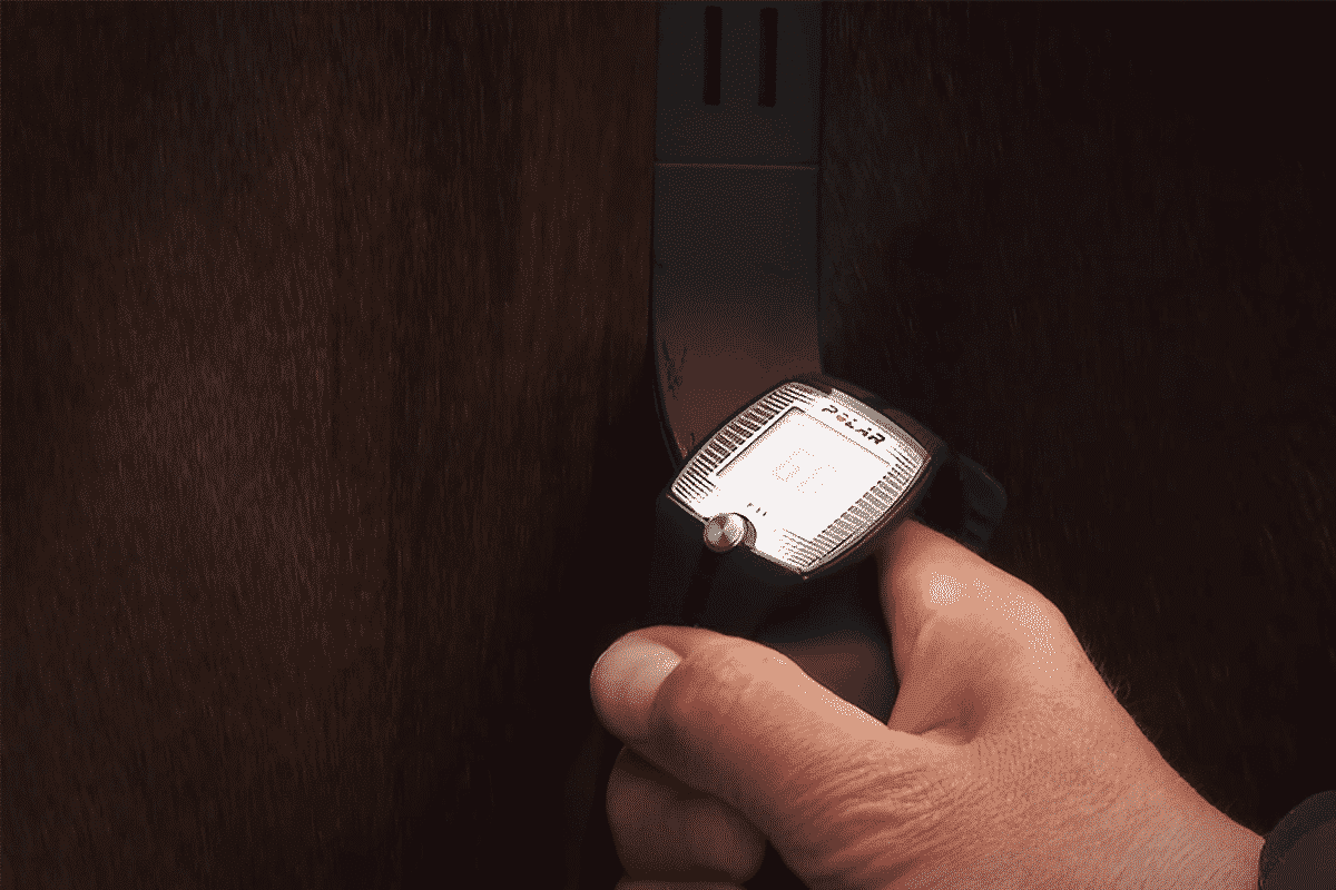
Heart rate monitors work similarly to ECGs in that they detect and measure the electrical activity produced by the heart, that is, the electrical voltage of the QRS wave. The HR monitor band has 2 inbuilt electrodes and the time duration between these larger voltage waves is measured over a few cycles and then averaged. The band is attached to a handle bar which should be placed firmly against the skin above the near side girth area in a vertical position. The area should be wet or even have electrode gel applied. The monitor band also contains a transmitter which sends the information to a display, usually a special watch. The display shows your horse’s heart rate. The transmitter and receiver are usually coded to ensure that signals are only between each unit and the watch is usually required to be within 1 m of the transmitter. Heart rate monitors can be used to assess resting HR’s , Post exercise HR’s , and pre vetting HR’s in the endurance setting, but can also be used to measure the HR during training and competition using electrodes attached under the girth and/or saddle. The advantage of the HR monitor over the stethoscope is that time to get an initial HR is much quicker as you are not counting the full minute, and you can easily and quickly check that the HR is consistently dropping.
How to use a HR Monitor (tips):
- Clean the electrodes on the band strap
- Read the instructions
- Make sure the skin is very wet or use gel
- Make sure the watch battery is not low (can be correctly replaced by Polar Electro Australia)
- Practice using the monitor at home, some horses find it uncomfortable at first
- Know how your horses HR’s change post exercise, especially in the extremes of heat and cold
- Know how your horses HR changes with eating, excitement, anxiety, walking, urinating, whinnying
- Know your HR cut-offs for the event you are participating in e.g. AERA training ride v 80km first leg, second leg versus vet gate into hold (VGIH) and FEI.
- Practice so that you know how low the HR needs to be so that you are “safe” to go to vetting in early vetting and VGIH situations.
- Have a stethoscope ready as a backup just in case, or even a second monitor.
- Label your HR monitor in permanent pen and also with bright coloured tape or the like to make it easily identifiable should you accidently part with it in a busy crewing area!
- Always listen to the heart with a stethoscope at some point as well, in order to detect any irregularities.
Troubleshooting
- Occasionally the watch monitor will read a high HR which is actually double the correct HR. This is due to the band electrodes picking up the electrical voltages from both the large QRS wave and the T wave. The sensitivity of most ECG equipment and HR monitors is set for humans, thus counting the high T wave is a common finding. Turning the band around 180 degrees and therefore swapping the electrode position, may change the voltages enough to correct this problem. Check with a stethoscope. Often as the HR slows even further, it will display the correct HR.
- The monitor can sometimes take longer than normal to display a HR. This is usually due to lack of moisture or poor contact electrode contact because the band is too low and the electrodes and not sitting flat on the horse.
In summary, the HR monitor is a very useful tool for speeding up the strapping process at an endurance ride. The HR is obtained by a different physiological process to using the stethoscope and the instantaneous HR is able to be measured thereby allowing a better insight into how the horse is recovering from the endurance activity being asked of it.

